
A seemingly minor twist of the foot can sometimes mask a devastating injury. This guide explores the Lisfranc complex, from the moment of injury to the best shoes for lifelong protection.
What Exactly Is a Lisfranc Fracture?
A Lisfranc fracture is more than just a broken bone — it is a disruption of the critical articulation between the midfoot and the forefoot. Named after French surgeon Jacques Lisfranc, this injury involves the metatarsal bones and their connecting ligaments that form the arch of your foot. When this structure is compromised, the stability of the entire foot is threatened.
Unlike a simple toe fracture, a Lisfranc injury is a fracture-dislocation of the tarsometatarsal (TMT) joints. It ranges from a subtle ligament sprain to a complete dislocation with multiple broken bones. The hallmark of the injury is instability, which often leads to long-term arthritis if not treated appropriately.
Anatomy of the Lisfranc Complex — Why this joint is unique
The Lisfranc complex is the keystone of your midfoot. It consists of the five metatarsal bones, the three cuneiforms, and the cuboid bone. The Lisfranc ligament itself runs from the medial cuneiform to the base of the second metatarsal — it is the only ligament that connects these two specific columns of the foot.
This ligament is essential because the second metatarsal is recessed into the tarsal bones, creating a “mortise and tenon” joint similar to a carpenter’s joint. This design provides intrinsic stability to the arch. When the Lisfranc ligament tears, the arch collapses, leading to a flatfoot deformity and severe functional impairment.
How Does a Lisfranc Injury Happen?
Lisfranc fractures occur in two distinct settings: high-energy trauma and low-energy sports injuries. Understanding the mechanism of injury is crucial for first responders and patients alike, as the force required to disrupt this joint is often underestimated.
What are the most common mechanisms of a Lisfranc injury?
High-Energy Trauma — Crush & axial load
Motor vehicle accidents and falls from height account for the majority of severe Lisfranc fracture-dislocations. In these scenarios, the foot is often plantarflexed (pointed down) while a crushing force is applied to the heel. The result is a catastrophic disruption of the midfoot architecture, frequently involving multiple metatarsal fractures and dislocations.
These injuries are usually obvious on X-ray because of the severe displacement. Patients often have other traumatic injuries that take priority, but the foot injury should never be overlooked.
Low-Energy Sports Injury — The “kissing injury”
In athletes, the Lisfranc joint is often injured during a forced plantarflexion and rotation event. Think of a football player whose foot is planted while another player falls onto the back of their heel. The same mechanism occurs in soccer, surfing, and cycling.
These low-energy injuries are the ones that are frequently missed. The displacement may be subtle, and the patient may walk off the field thinking they just have a “bad sprain.” However, the Lisfranc ligament can be completely ruptured without a single bone breaking.
Simple Falls & Missteps — The curb stone injury
Even a simple misstep off a curb can cause a Lisfranc injury, particularly in older adults or those with osteopenia. As the foot rolls into extreme plantarflexion, the full body weight is transmitted through the TMT joints. The resulting injury can be subtle, with only a small fleck of bone avulsed from the ligament insertion point.
This is the classic “missed” Lisfranc injury. Because the bones may look aligned on a non-weight-bearing X-ray, the injury is dismissed as a sprain. It is only when the patient returns weeks later with persistent pain that a weight-bearing X-ray reveals the truth.
Symptoms & Red Flag Warning Signs
Recognizing a Lisfranc injury early is the single most important factor in achieving a good outcome. The symptoms can be subtle, but there are several classic red flags that differentiate this injury from a simple ankle twist.
What does a Lisfranc fracture feel like?
The most consistent symptom is pain over the top of the midfoot, specifically between the arch and the toes. Swelling is universal, but the location of the swelling and bruising is what makes Lisfranc injuries unique.
If you have sustained any injury to your foot and you cannot walk more than four steps, you need an X-ray. Do not simply “walk it off.” A delay in diagnosis of a Lisfranc injury leads to higher rates of post-traumatic arthritis and the need for complex reconstructive surgery.
The Critical Misdiagnosis: Myths vs. Reality
Lisfranc injuries are among the most commonly missed foot injuries in emergency medicine. The reason is simple: the bones may appear perfectly aligned on a standard X-ray if the patient is not standing. Here, we debunk the most persistent myths about this challenging diagnosis.
False. Non-weight-bearing X-rays miss 20-40% of Lisfranc injuries. Weight-bearing X-rays or CT scans are often required to see the subtle separation of the metatarsals. A “normal” X-ray does not rule out a Lisfranc ligament injury.
Partially False. Non-displaced fractures and stable ligament injuries can be treated without surgery. However, any instability or widening of the joint usually requires surgery to restore the anatomy. The decision depends on whether the joint is stable.
True, but manageable. Even with perfect surgical reduction, the cartilage damage at the time of injury is often irreversible. Up to 50% of patients develop post-traumatic arthritis. However, proper treatment and footwear can significantly reduce pain and maintain function.
| Feature | Lisfranc Injury | Simple Midfoot Sprain |
|---|---|---|
| Location of Pain | Over the TMT joints (dorsal midfoot) | Over ligaments (diffuse) |
| Plantar Bruising | Strongly indicative | Very rare |
| Weight Bearing | Excruciating / Impossible | Painful but possible |
| X-ray Findings | Diastasis, fleck sign, fracture | Usually negative |
| Long-term Prognosis | High risk of arthritis | Excellent |
Treatment: Surgical vs. Non-Surgical Paths
The treatment of a Lisfranc fracture is determined by one factor: stability. If the joint is stable and the bones are perfectly aligned, non-surgical treatment may work. If there is any displacement or instability, surgery is the gold standard to prevent catastrophic arthritis.
Indication: Non-displaced, stable fractures.
Protocol: 6-8 weeks in a non-weight-bearing cast, followed by 4 weeks in a walking boot. Frequent X-rays are needed to ensure no late displacement.
Outcome: Good, provided the ligament is intact and the bones remain aligned.
Indication: Any displacement, dislocation, or instability.
Protocol: Open Reduction and Internal Fixation (ORIF) with plates and screws. In some cases, Primary Arthrodesis (fusion) is performed for severe cartilage damage.
Outcome: Anatomic alignment is restored. Hardware removal is often needed later.
What is the best surgery for a Lisfranc fracture?
The decision between ORIF and Primary Arthrodesis is debated among foot and ankle surgeons. ORIF preserves joint motion but requires a second surgery to remove the screws. Arthrodesis fuses the joint, eliminating motion at that articulation but providing a stable, pain-free weight-bearing surface. Recent studies suggest that for purely ligamentous injuries, ORIF has a slight advantage in maintaining foot flexibility.
“The Lisfranc joint is structurally akin to a Roman arch — each stone relies on its neighbor for stability. When the keystone ligament fails, the entire arch collapses. Surgery is about rebuilding that arch.”
— Dr. David Thurman, Orthopedic Foot Specialist
Recovery Timeline & Physical Therapy
Recovery from a Lisfranc injury is a marathon, not a sprint. Patients must be prepared for a prolonged period of non-weight-bearing and a gradual return to full activity. Rushing this process can lead to hardware failure or chronic pain.
How long does it take to recover from a Lisfranc fracture surgery?
The typical recovery is 4 to 6 months for return to daily life, and up to 12 months for return to high-impact sports. Here is the standard rehabilitation roadmap used by physical therapists in 2026.
Patients should expect some degree of long-term stiffness and swelling. Night pain and first-morning pain are common for up to a year. Proper footwear becomes a permanent part of managing foot health post-Lisfranc injury.
Best Shoes for Lisfranc Joint Protection
After a Lisfranc injury, your shoes are your primary defense against re-injury and arthritis progression. The right shoe minimizes motion through the TMT joints, provides arch support, and reduces the bending force on the midfoot. Flexible shoes are the enemy of the Lisfranc joint.
What features should you look for in a shoe after a Lisfranc fracture?
- Hoka Bondi 8 / Clifton 9: Excellent rocker sole, very stiff, maximum cushion.
- ASICS Gel-Kayano 30: Best in class stability, secure lacing system.
- Brooks Addiction Walker: Firm sole, great for daily walking, durable.
- Vionic Uptown Sneaker: Structured orthotic footbed, firm heel counter.
- Mojo Sport Carbon: Specific carbon fiber plate shoe for post-surgical recovery.
Avoid: Minimalist shoes, barefoot-style sneakers, flip-flops, or any shoe that you can easily fold in half. These shoes force the Lisfranc joint to absorb all the bending forces of gait.
Critical Mistakes to Avoid During Healing
Even with perfect surgery and a great PT protocol, patients can sabotage their recovery by making a few common errors. Here is what every Lisfranc patient should avoid.
If you are returning to high-impact sports, consider using a Lisfranc-specific carbon fiber orthotic inside your cleats or athletic shoes. Brands like OrthoGait and Bledsoe make custom plates that bolt into the shoe to prevent midfoot bending during cutting and jumping.
Frequently Asked Questions (FAQ)
Here are the most common questions we receive from patients navigating a Lisfranc fracture diagnosis and recovery.
Is a Lisfranc fracture a break or a sprain?
It is both. The term “Lisfranc fracture” encompasses a spectrum of injuries from a purely ligamentous tear (sprain) to a fracture-dislocation. Because the ligament is the primary stabilizer, a “Lisfranc sprain” is often more functionally disabling than a simple fracture because it destabilizes the arch.
Can you walk on a Lisfranc fracture?
Technically, some people can walk, especially if it is a subtle injury. However, walking on a displaced Lisfranc injury will cause the arch to collapse over time. If you have a confirmed Lisfranc injury, you should be non-weight-bearing until cleared by your surgeon. Walking on it worsens the deformity.
How long does it take to walk normally after surgery?
Most patients are in a non-weight-bearing cast for 8 to 12 weeks. After that, they transition to a walking boot for 4 to 6 weeks. You can expect to walk somewhat normally in a supportive shoe by the 4th month, but a limp may persist for 6 to 12 months due to stiffness and weakness.
What happens if a Lisfranc injury is left untreated?
Untreated Lisfranc instability leads to post-traumatic arthritis, a painful collapse of the midfoot arch, and a progressive flatfoot deformity. This often necessitates a complex reconstruction or fusion surgery years later, which is more invasive than the initial treatment would have been.
Will I need surgery to remove the hardware?
Often, yes. The screws used to stabilize the Lisfranc joint cross the TMT joints. Once the ligaments have healed (usually at 4-6 months), the screws are removed to restore motion at those joints. Hardware removal is usually a minor outpatient procedure with a quick recovery.
The Bottom Line on Lisfranc Fractures in 2026
A Lisfranc fracture is a serious injury that demands respect. The single most important message from this guide is this: do not let a midfoot injury be dismissed as a “sprain” without proper weight-bearing imaging. The window for optimal treatment is narrow, and early diagnosis is the best predictor of a good outcome.
Modern surgical techniques offer excellent results for restoring anatomy, and advances in physical therapy help patients regain strength and mobility faster than ever before. However, the patient’s commitment to the non-weight-bearing phase and to making permanent footwear upgrades is what ultimately determines long-term success.
“The Lisfranc injury is a humbling condition. It forces athletes and active individuals to slow down, comply with strict protocols, and accept that the foot needs time. Those who do well are the ones who respect the process.”
— Orthopedic Rehabilitation Protocol, Johns Hopkins Medicine
Final recommendation: If you suspect a Lisfranc injury, see a foot and ankle orthopedist immediately. Bring this guide to your appointment to ask specific questions about stability, weight-bearing status, and the best type of shoe for your specific injury pattern.
You may also like
-
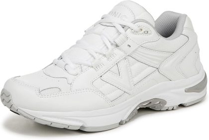
Vionic Women’s 23walk 2.0
$110.00 -
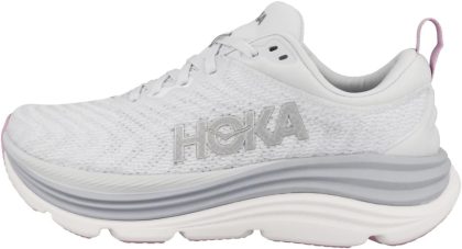
Hoka ONE ONE Womens Gaviota 5
$196.89 -
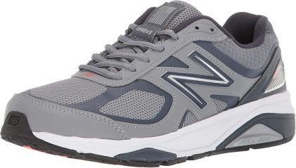
New Balance Women’s Made in US 1540 V3 Running Shoe
$119.99 -

ASICS Women’s Gel-Kayano 31 Running Shoes
$124.95